
Healthbeauty123.com – The symptoms of a clot in the Vertebral Basilar Artery are similar to those of cerebral artery occlusion. In some cases, the patient may even lose their balance or drop their arms, which are often signs of an ischemic stroke. A positive test may also be accompanied by sensory changes or nystagmus. In severe cases, thrombolytic therapy is used to relieve the clot.
Causes Permanent Brain Damage and Long Term Treatment
If the blood flow to the brain is restricted, the patient will experience various symptoms. These symptoms will vary from person to person and the extent of the restriction. Medications that lower cholesterol levels and control platelet function are commonly used. If diagnosed early, patients may be able to get a treatment that will improve their quality of life. If left untreated, the disease may lead to permanent brain damage and long-term care.
There are several causes of reduced blood flow to the back of the brain. Smoking, high blood pressure, high cholesterol, and diabetes are the most common risk factors. If you suspect that you have this condition, it’s vital to see a vascular surgeon. While these procedures are not proven to cure this problem, they can help reduce your symptoms. Depending on your situation, you may need a medical intervention to fix the problem.
The findings of the study suggest that stenoses in the vertebral arteries may be risk factors for basilar artery disease. According to the authors of the study, the presence of stenoses in the vertebral artery could be a risk factor for developing this condition. Moreover, it is important to note that a stenosis in the vertebral artery may be a factor in basilar artery disease.
High Risk of Basilar Artery Disease
Infarcts of the circulation are responsible for up to 12-27% of all strokes in hospitalized patients. These stenoses are typically classified according to their location and underlying cause. There are a variety of other causes of the disease. Some of these may include cardiac disease, ischemic heart conditions, and pulmonary arterial hypertension. Despite the high risk of basilar artery disease, it can be treated successfully with medications and surgery.
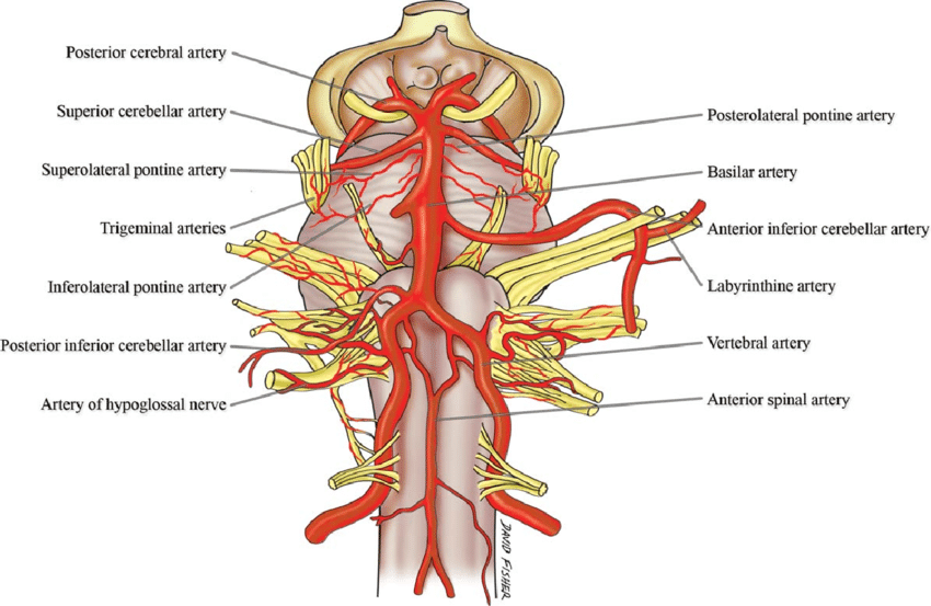
The vertebral Basilar artery is an artery in the vertebral column of the brain. It supplies the cerebellum, the OCCIPITAL LOBE, and the THALAMUS. If the arteries are blocked, the symptoms of the disease will be the same as those of a stenosis in the Cervical arteries. If the stenosis is in the Vertebral Basilar Artery, the underlying condition is known as vertebrobasilar insufficiency or vertebral artery stenosis.
A stenotic Vertebral Basilar Artery can result from a number of underlying causes. Acute stroke can be diagnosed by a basic blood test. Usually, the arteries are analyzed in a CT scan to detect the type of disease. This procedure is performed in the operating room under general anesthesia. A stenosed Vertebral Basilar Artery should be immediately removed.
Blocked Carotid Artery Increases Stroke Risk
The vertebral basilar artery is one of the most important blood vessels in the body. It is located in the neck on both sides and carries blood to certain parts of the brain. A blocked carotid artery increases the risk of a stroke. A swollen Vertebral Basilar Artery is a serious medical condition that causes pain and disability. The disease can lead to stroke and even death.
There are two subgroups of basilar arteries: unilateral and bilateral. The former has larger diameter than the latter. The latter is characterized by asymmetrical morphology and is the most common type of stenosis in the Vertebral Basilar Artery. The left VA is typically larger than the right, whereas the right VA is frequently smaller than the right. Regardless of the sclerosis, the underlying anatomical differences in the Vertebral Basilar artery are unknown.
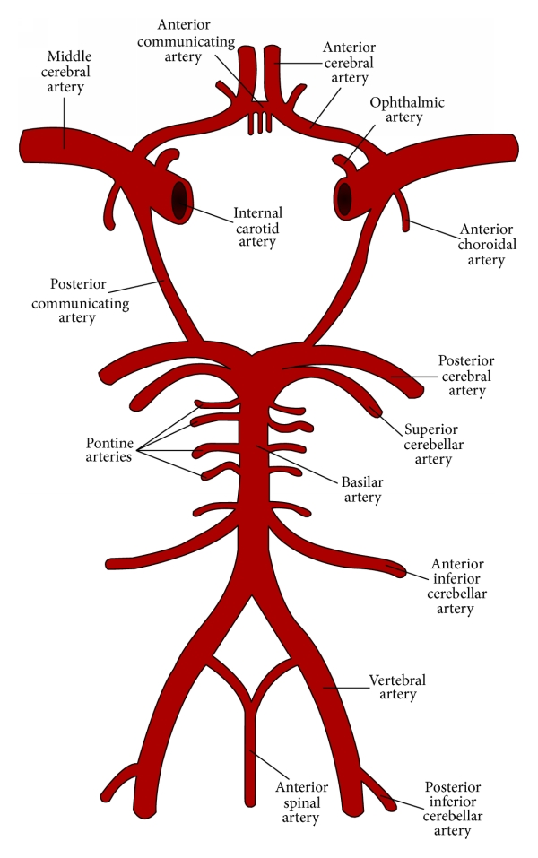
In acute strokes, the Vertebral Basilar Artery is the most common cause of a stroke. It supplies both the posteroinferior and the posterior hemispheres of the cerebellum. The basilar artery is divided into two posterior and superior cerebral arteries. At the proximal midbrain, the vertebral basilar artery and the thoracic arteries meet and merge.
Reference:


