Healthbeauty123.com – A common cause of arthritis in the thumb is a torn beak ligament. This injury can result from a variety of causes, including trauma or a degenerative disorder. In order to understand the most effective treatment, a thorough diagnosis of the affliction is necessary. This article will explain the most common methods of treating this disorder and what your options are. A proper diagnosis is important to avoid complications later.
Surgical Reinforcement of the Beak Ligaments
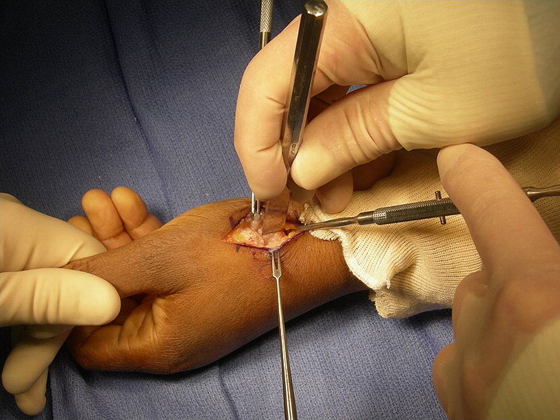
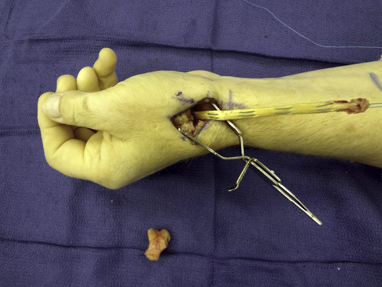
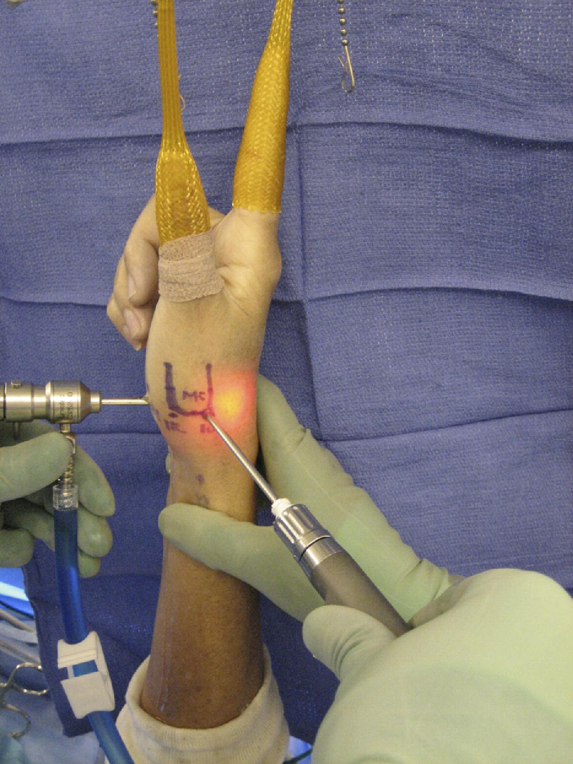
The first method involves a high index of suspicion and a thorough clinical history. Surgical re-enforcement of the beak ligament is important to restoring global stability to the thumb. It is best performed by a surgeon experienced in hand surgery. A case report of this operation was written by Mr. Karthikeyan Iyengar, the author of the article, and Mr. Jayant Nadkarni, the second author of the study. A CT-derived bone model and joint kinematics are used to compute the length of the beak ligament. These models provide an insight into the elongation of the ligament and the mechanism of its rupture.
The AOL is a thick ligament in the hand. It is much thinner than the dorsal ligament complex. It is functionally important and is the part that pulls in the radial direction. The AOL and APL pull together in a wide diastasis. Although the superficial AOL is not completely visible, the striated appearance of the deep AOL makes differentiation difficult. X-ray control of the scaphoidal joint and the volar beak ligament is essential in the diagnosis of this disorder.

The clinical diagnosis of this condition depends on a patient’s symptoms, age, and type of OA. ACT-based bone model can help determine the cause of the affliction. In vivo measurements of the ligament’s length are impractical. But computational approximations of ligament elongation can be used. This technique provides an understanding of how the ligament supports the joint and its pathomechanics.
Evaluating Beak Ligament Area
Radiographs of the beak ligament are important for assessing the cause of the problem. A plain radiograph will show if the CMC joint is dislocated. A lateralized beak ligament can also result in a shoulder sign. The CMC joint is commonly affected in both OA and RA. However, a careful evaluation is necessary for a thorough diagnosis. In a cadaveric patient, a CT-derived bone model can be used to evaluate the extent of the beak ligament.
A CT-derived model of the beak ligament can be used to determine its length. Using the CT-derived bone model, a surgeon can identify OA and its causes. They will perform a CT-based study to assess the severity of the disease and to develop an intervention plan. The patient will be treated according to his or her unique needs. The OA treatment will depend on the type of underlying problem.

A study published in the Journal of Hand Surgery, Inc., showed that the ligament was responsible for OA. The researchers found that OA caused the AOL to tear. The x-rays were not able to confirm the cause of OA. But they were able to demonstrate the damage with a CT-derived image. This is an example of the use of a CT-derived image to diagnose the afflicted area.
Confirming the Presence of an Elongated Beak Ligament
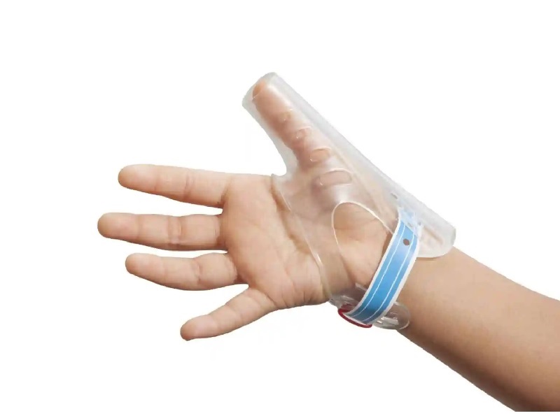
The CT-based model was used to measure the length of the beak ligament. They were able to confirm the presence of an elongated beak ligament in patients with OA. The MRI results were correlated with OA. After the surgery, the patient was placed in a scaphoid cast for four weeks. The k-wires were removed after six weeks. The patient did not respond to the treatment, and failed to undergo provocation stress tests. In addition, the x-ray control confirmed the presence of a ruptured volar beak ligament.

The study was published in JHS, where the authors described the treatment of a patient with a torn beak ligament. It was followed by a CT-derived bone model. The MRI was performed to verify the underlying cause. The anatomical illustrations were made by Miss Priya Gandham and her colleagues. This research revealed that beak ligaments could cause the pain of the thumb and the articular elongated beak.
Reference:


