
Healthbeauty123.com – Direct trauma to the extensor tendon sheath causes swelling in the carpus and hock. The pattern of swelling varies depending on the exact location of the injury. Acute injury to the tendon itself within the sheath can cause chronic swelling and lameness. In order to diagnose this condition, a veterinarian will first perform an ultrasound of the joint, and sample tendon sheath fluid if necessary.
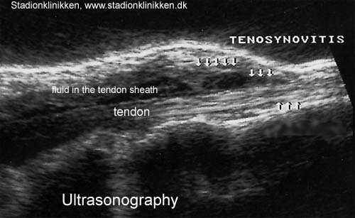
Ultrasound can Help Visualize the Extensor Tendon Sheath
Axial T2-weighted fat-suppressed MR images show fluid in the extensor tendon sheaths, and adjacent soft-tissue edema is present in the proximal region. The presence of fluid within the tendon sheath indicates tenosynovitis. In some cases, the condition may be associated with bone marrow edema, which may be present in the radius.
An ultrasound can help visualize the extensor tendon sheath. The distal third of the foot is scanned, and MRI can show the integrity of cortical bone and the extent of effusion and hypertrophy of the tendon sheath. The distal third of the foot is also analyzed using MRI to determine if the tendon sheath has ruptured or not.
Surgery may be required in severe cases of tendon inflammation. The surgical procedure may repair the joint and allow the tendon sheath to heal. In addition to surgery, a rehabilitation program may be recommended to help patients regain strength and learn how to prevent further injuries. If necessary, the patient will receive physical therapy and occupational therapy to help prevent further damage. These treatments may include strengthening the muscles around the joint. Further, they can help patients develop the ability to avoid injure themselves in the future.
The Lesion is Located within the Extensor Tendon Sheath
A 30-year-old woman presented with a swelling in her right thumb for three months. The swelling was slow-growing, ill-defined, and mobile only in the vertical plane. Diagnostic X-ray showed slight erosion of the first metacarpal head, and a diagnostic ultrasound confirmed that the lesion was located within the extensor tendon sheath. A wide local excision biopsy was planned.
Giant cell tumors of the tendon sheath are rare, and are often benign. Most often found in the hand, they can also occur in the foot and ankle. They are most common in younger adults and often accentuate deformities of the toe. Treatment involves surgery. This condition is usually treated surgically, but may require a radiotherapy procedure if it returns. When treatment fails, surgery may be required.
An MRI revealed that the EPL and EPRL tendons share a common sheath prior to their divergence. The ECRL and ECR tendons diverged at the distal end of the EPL ten don. The contrast material flowed without obstruction in either tendon sheath. This condition was diagnosed in a skier and kickboxer. The patient had pain in the radial dorsal wrist region.
Experiencing Pain and Swelling in the Ulnar Wrist Back
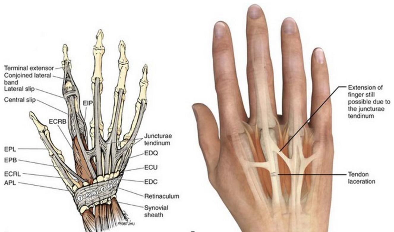
The ECU is located in the wrist where it courses across the distal ulna. The ECU sheath is protected by a fibro osseous tunnel. The volar aspect of the ECU subsheath is confluent with the distal radioulnar joint capsule and the dorsal radioulnar ligation. The ECU subsheath stabilizes the tendon as it travels over the distal ulna. Located deep to the extensor retinaculum, it prevents the tendon from bowstringing. Patients with ECU subsheath injuries commonly have pain and swelling on the dorsal ulnar wrist. This pain is exacerbated by ulnar deviation.
Treatment for EPL tendon inflammation depends on the cause of the problem and its duration. Generally, nonsteroidal anti-inflammatory drugs or injectable corticosteroids will alleviate the inflammation. If it is caused by an infection, however, a surgical procedure may be necessary to release the tendon sheath. On the other hand, antibiotics will treat the infection. The resulting inflammation will usually clear up after a few days.

In some cases, a patient may require surgery to remove the thickened fibro-osseous sheath surrounding the extensor tendons in the wrist. This procedure can be performed endoscopically or openly and is considered elective outpatient surgery. This procedure is often necessary after conservative therapies such as nonsteroidal anti-inflammatory drugs, splinting, or activity modification. However, the risk of rapid wrist deviation and pain should not discourage patients from undergoing this procedure.
Reference:


