
Healthbeauty123.com – The plantar sesamoid bone is a small joint located within the fibrous plantar pad. Its main function is to distribute and absorb weight from the head of the metatarsal. They also increase the mechanical advantage of motion at the first MPJ and the moment arm of the flexion muscles. During walking, running, and jumping, the sesamoid is an important structural component. But how does it function?
Sesamoids Gradually Grow in Size as a Child Grows
The first appearance of the plantar sesamoid occurs 1.35 years before the onset of PHV. In contrast, the lateral radiograph shows sesamoids prior to the onset of the disease. The two types of sesamoid are usually evident at a young age. They develop in similar periods. They first appear on the foot at a relatively young age, while they gradually grow in size as a child grows.
Most dogs do not experience lameness due to sesamoid disease, although some will experience pain when the joint is manipulated. Later on, the condition can progress to osteoarthritis, a condition characterized by poor range of motion and thickening of the joint. The front foot is more often affected. Diagnosis is usually made through X-rays, although anesthesia is necessary to visualize the sesamoid bone in “stress” positions.
In cases of complete absence of sesamoids, doctors may opt to preserve the entire plantar sesamoid or repair the DP. If there is an asymptomatic sesamoid, the remaining sesamoid bone may be excised and soft tissues repaired through drill holes. The remaining soft tissue defect may be large and compromise flexor power. A partial sesamoid may also cause a fixed deformity. If the sesamoid is reconstructed, the abductor hallucis tendon can be transferred plantarly to provide structural support and collagen.
Sesamoid Hand Injury Can Have Substantial Effects
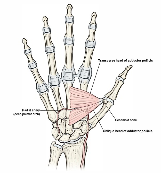
In addition to the foot, the hand has sesamoid bones. In most people, there are five sesamoids in the hand. The most common ones are the two sesamoids located at the base of the thumb and the distal aspect of the first metacarpal. Sesamoid bones are easily overlooked, but they play a significant role in hand and foot injuries. Hand sesamoid injury can have a substantial effect on a person’s daily life.
If the sesamoid bone is displaced, it may be a sign of a plantar plate rupture. Sesamoids should not migrate with the range of motion of the first MTP joint. If they migrate to the lateral side, the sesamoid is highly likely to rupture. A distal sesamoid-to-joint distance greater than 10 mm is highly suggestive of a rupture of the plantar plate.
In addition to running, the increased popularity of athletic activities like cycling and running has made sesamoid insufficiency a common condition in the foot. Foot surgeons must be familiar with sesamoid anatomical variations and their symptoms. The early recognition of sesamoid insufficiency allows for diagnosis and treatment that prevents long-term complications. So, how can we know whether we are experiencing a sesamoid insufficiency?
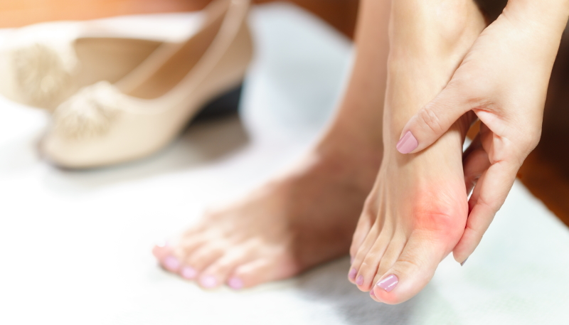
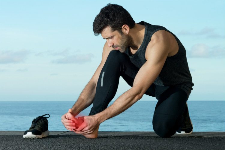
The First Symptoms Associated with a Sesamoid Pantar Injury
The first symptoms associated with plantar sesamoid injury are aching and pain in the first metatarsal head. Pain in walking, running, and jumping may also be present. However, in many cases, pain is relieved by increasing weight bearing. The condition may also result in stiffness or a fracture of the sesamoid bone. To treat a sesamoid injury, it is important to determine the cause of the pain.
During surgery, a second metatarsal is exposed from the medial perspective. The first metatarsal is removed and the second one is placed in its place. The first dorsal interosseous muscle may need to be divided. Sometimes, an intramuscular dissection is necessary to expose the dominant inflow system. These procedures may be painful and require a lengthy recovery period. But the results will be worth it.
The foot and ankle complex is made up of the tibia, fibula, talus, and calcaneus. In addition to the sesamoid bone, the foot and ankle complex is comprised of six ligaments and fascicles. The tibia and fibula form the hindfoot. The calcaneus and talus are connected by a subtalar joint.


